Smooth Muscle Diagram Labeled : Smooth Muscle Wikipedia : Drag the labels onto the diagram to label the steps of smooth muscle activation and deactivation.
Smooth Muscle Diagram Labeled : Smooth Muscle Wikipedia : Drag the labels onto the diagram to label the steps of smooth muscle activation and deactivation.. Fill in all 8 boxes. Similar to skeletal muscle tissue, cardiac muscle does not regenerate to a great extent. It constitutes much of the musculature of The calcium is the cause of protein to detach from the actin and myosin fastly binds with the opening of actin. Kierszenbaum, al histology and cell biology 2nd ed., mosby elsevier, 2007, p.
The diagram is fully labeled. Muscle fibers are organized into bundles supplied by blood vessels and innervated by motor neurons. Related posts of smooth muscle diagram labeled muscle anatomy crossword key biology corner. Thousands, or even tens of thousands, of small fibers make up each muscle. In skeletal muscle, a single type of somatic nervous system traverses to muscle, where it stimulates organelle in the muscle cells in order to release calcium.
Differentially Expressed Proteins Visualized In The Pathway Of Vascular Download Scientific Diagram from www.researchgate.net Pericytes allow smooth muscle cells to regenerate and repair much more readily than skeletal and cardiac muscle tissue. Skeletal muscle tissue is composed of long cells called muscle fibers that have a striated appearance. Smooth muscle (textus muscularis levis) smooth muscle is a type of tissue found in the walls of hollow organs, such as the intestines, uterus and stomach. Anatomy of outer ear diagram. This diagram shows a few of the cells that can be seen in the stained section below. The human body has three different types of muscles. Drag the labels onto the diagram to label the steps of smooth muscle activation and deactivation. Drag the labels onto the diagram to label the steps of smooth muscle activation and deactivation.
Termed unitary smooth muscle or visceral muscle, this type of smooth muscle is the most common observed in the human body, forming the walls of hollow organs.
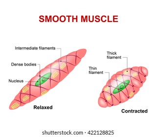
Smooth muscle tissue can regenerate from a type of stem cell called a pericyte, which is found in some small blood vessels. Skeletal muscle tissue is composed of long cells called muscle fibers that have a striated appearance. A gap junction (gj) is present in the longitudinal portion of the disk. Pericytes allow smooth muscle cells to regenerate and repair much more readily than skeletal and cardiac muscle tissue. They range from about 30 to 200 μm (thousands of times shorter than skeletal muscle fibers), and they produce their own connective tissue, endomysium.although they do not have striations and sarcomeres, smooth muscle fibers do have actin and myosin. Thousands, or even tens of thousands, of small fibers make up each muscle. Neuromuscular junctions of the highly structured type found on skeletal muscle fibers do not occur in smooth muscle. Muscle anatomy back 12 photos of the muscle anatomy back back muscle anatomy images, back muscle anatomy of the human body, back pain muscle anatomy, muscle anatomy lower back, posterior back muscle anatomy, human muscles, back muscle anatomy images, back muscle anatomy of the human body, back pain muscle anatomy, muscle. It is the pen diagram of skeletal, smooth and cardiac muscle for class 10, 11 and 12. Smooth muscle cells are found in the dividers of empty organs, including the stomach, digestion tracts, urinary bladder and uterus, and in the dividers of paths, for example, the supply routes and veins of the circulatory framework, and the tracts of the respiratory, urinary, and regenerative frameworks. The calcium is the cause of protein to detach from the actin and myosin fastly binds with the opening of actin. Physiologic anatomy of smooth muscle neuromuscular junctions. In this short guide, you will get a basic concept of skeletal muscle histology from the real slide and labeled diagram.
Smooth muscle, muscle that shows no cross stripes under microscopic magnification. The human body has three different types of muscles. Smooth muscle (textus muscularis levis) smooth muscle is a type of tissue found in the walls of hollow organs, such as the intestines, uterus and stomach. Smooth muscle fibers are often found forming sheets of tissue and function in a coordinated fashion due to the presence of gap junctions between the cells. Human muscles · july 29, 2021.
Smooth Muscle Hd Stock Images Shutterstock from image.shutterstock.com In this video i have shown the simplest way of drawing muscle drawing. 12 photos of the smooth muscle diagram. Physiologic anatomy of smooth muscle neuromuscular junctions. Muscle anatomy back 12 photos of the muscle anatomy back back muscle anatomy images, back muscle anatomy of the human body, back pain muscle anatomy, muscle anatomy lower back, posterior back muscle anatomy, human muscles, back muscle anatomy images, back muscle anatomy of the human body, back pain muscle anatomy, muscle. You have three different types of muscles in your body: Smooth muscle is made up of cells that contain a single central nucleus. The smooth muscles perform the functions in the contrast of other types of muscles. The cells are spindle shaped, and the nucleus is central.
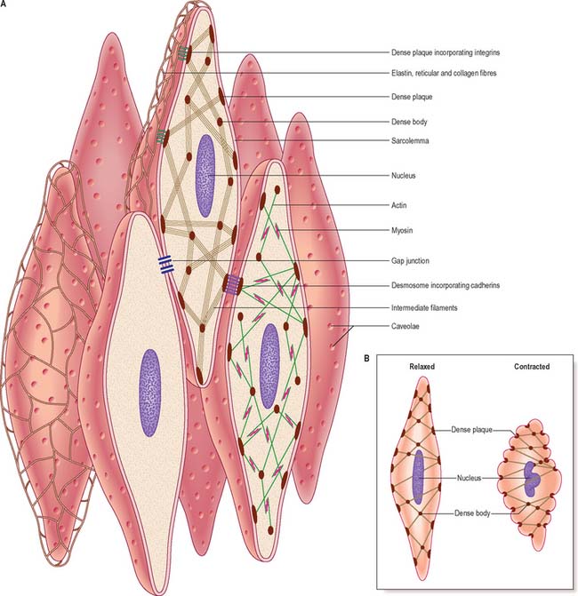
The cells stick together and are connected by specialised cell junctions, called gap junctions.
In skeletal muscle, a single type of somatic nervous system traverses to muscle, where it stimulates organelle in the muscle cells in order to release calcium. The cells stick together and are connected by specialised cell junctions, called gap junctions. Muscles are all made of the same material, a type of elastic tissue (sort of like the material in a rubber band). Termed unitary smooth muscle or visceral muscle, this type of smooth muscle is the most common observed in the human body, forming the walls of hollow organs. Smooth muscle tissue smooth muscle cell smooth muscle cells types muscles skeletal muscle skeletal muscle cell muscle cell heart muscle cells pictures of muscles smooth muscle contraction. Skeletal muscles, smooth muscles, cardiac muscles. Related posts of smooth muscle labelled diagram muscle anatomy back. In skeletal muscle, a single type of somatic nervous system traverses to muscle, where it stimulates organelle in the muscle cells in order to release calcium. Kierszenbaum, al histology and cell biology 2nd ed., mosby elsevier, 2007, p. Muscle anatomy crossword key biology corner 12 photos of the muscle anatomy crossword key biology corner muscle anatomy crossword answer key biology corner, muscle anatomy crossword key biology corner, muscle anatomy crossword puzzle answers biology corner, human muscles, muscle anatomy crossword. You will also get the identification points of skeletal muscle histology slide with a little description here in this guide. Smooth muscles are unique in their largely involuntary response, and in their structure. Thousands, or even tens of thousands, of small fibers make up each muscle.
Fill in all 8 boxes. The muscles of the human body can be categorized into a number of groups which include muscles relating to the head and neck, muscles of the torso or trunk, muscles of the upper limbs, and muscles of the lower limbs. The skeletal muscle fibers are elongated, cylindrical and multinucleated cells whose length may vary in different animals. Muscle anatomy back 12 photos of the muscle anatomy back back muscle anatomy images, back muscle anatomy of the human body, back pain muscle anatomy, muscle anatomy lower back, posterior back muscle anatomy, human muscles, back muscle anatomy images, back muscle anatomy of the human body, back pain muscle anatomy, muscle. In skeletal muscle, a single type of somatic nervous system traverses to muscle, where it stimulates organelle in the muscle cells in order to release calcium.
Smooth Muscle And The Cardiovascular And Lymphatic Systems Clinical Gate from clinicalgate.com Smooth muscle, muscle that shows no cross stripes under microscopic magnification. The cells stick together and are connected by specialised cell junctions, called gap junctions. Smooth muscle tissue can regenerate from a type of stem cell called a pericyte, which is found in some small blood vessels. Smooth muscle fibers are often found forming sheets of tissue and function in a coordinated fashion due to the presence of gap junctions between the cells. In addition, the contractile state of smooth muscle is controlled by hormones, autocrine/paracrine agents, and other local chemical signals. This problem has been solved! Muscle anatomy back 12 photos of the muscle anatomy back back muscle anatomy images, back muscle anatomy of the human body, back pain muscle anatomy, muscle anatomy lower back, posterior back muscle anatomy, human muscles, back muscle anatomy images, back muscle anatomy of the human body, back pain muscle anatomy, muscle. Kierszenbaum, al histology and cell biology 2nd ed., mosby elsevier, 2007, p.
In this short guide, you will get a basic concept of skeletal muscle histology from the real slide and labeled diagram.
The smooth muscles perform the functions in the contrast of other types of muscles. Drag the labels onto the diagram to label the steps of smooth muscle activation and deactivation. Smooth muscle is made up of cells that contain a single central nucleus. Muscle fibers are organized into bundles supplied by blood vessels and innervated by motor neurons. Kierszenbaum, al histology and cell biology 2nd ed., mosby elsevier, 2007, p. Smooth muscle makes up the walls of hollow organs, respiratory passageways, and blood vessels. Pericytes allow smooth muscle cells to regenerate and repair much more readily than skeletal and cardiac muscle tissue. Similar to skeletal muscle tissue, cardiac muscle does not regenerate to a great extent. Drag the labels onto the diagram to label the steps of smooth muscle activation and deactivation. Human muscles · july 29, 2021. This diagram shows a few of the cells that can be seen in the stained section below. 12 photos of the smooth muscle diagram. This problem has been solved!
Muscle fibers are organized into bundles supplied by blood vessels and innervated by motor neurons smooth muscle diagram. Intercalated disk (ventricle, cat) click to see enlarged view:


0 Komentar